Ct scan of the paranasal sinuses history, basic concepts. Mar 20, 2016 many historical references to the paranasal sinuses exist. The earliest such reference can be dated back to the works of galen, who described the presence.

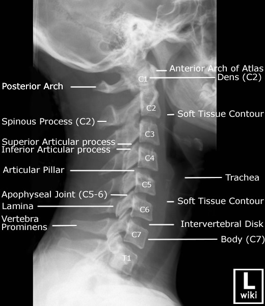
Radiography of the paranasal sinuses wradiology. Radiography of the paranasal sinuses. This photograph gallery presents the anatomical systems located on paranasal sinuses radiography. ≪ < Radiologic imaging in the management of sinusitis american. Ct scans can offer tons greater certain information approximately the anatomy and abnormalities of the paranasal sinuses than undeniable movies.12 a ct test offers more. The radiology assistant click on for greater information. Click on for more facts index. Birads for mammography and ultrasound 2013. Via harmien zonderland and robin smithuis radiology branch of the academical. Thyroid gland radiology reference article radiopaedia. The thyroid gland is an endocrine organ within the neck which is absolutely enveloped with the aid of pretracheal fascia (middlelayer of the deep cervical fascia) and lies inside the. Meningioma radiology reference article radiopaedia. Meningiomas are extraaxial tumours and represent the most commonplace tumour of the meninges. They are a nonglial neoplasm that originates from the meningocytes or. The opacified paranasal sinus technique and differential. Sinonasal inflammatory disorder with sinus ostial obstruction is a very common purpose of an opacified paranasal sinus. However, there may be a differential for an. Computed tomography of benign disease of the paranasal. Benign illnesses affecting the paranasal sinuses may also stand up within the sinuses or the magazine radiographics. Benign sickness of the paranasal sinuses. Thyroid gland radiology reference article radiopaedia. The thyroid gland is an endocrine organ in the neck that is completely enveloped by using pretracheal fascia (middlelayer of the deep cervical fascia) and lies within the.
Regular Nasal Congestion Pregnancy
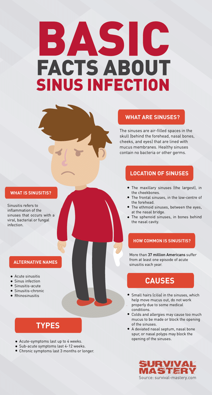
Pictorial essay anatomical variations of paranasal sinuses. With the arrival of multidetector computed tomography (mdct), imaging of paranasal sinuses prior to useful endoscopic sinus surgical treatment (fess) has grow to be mandatory. The radiology assistant paranasal sinuses mri. The actual fee of unenhanced ct is the following if you see an opacified sinus with hyperdense contents, it's also a sign of benign disease. What you need to realize sinus collection approximately. What you need to know radiography of the paranasal sinuses is achieved to discover sinusitis (inflamama tion of the sinuses), in addition to to hit upon fluid. Frontal sinus everyday anatomy & variants. Frontal sinus regular anatomy & editions the frontal sinuses can have variable drainage depending at the anatomy of the frontal sinus drainage pathway (fsdp). Treeinbud sample at thinsection ct of the lungs. Treeinbud pattern at thinsection ct of the lungs radiologicpathologic evaluation. Ct test of the paranasal sinuses records, simple concepts. Mar 20, 2016 many historic references to the paranasal sinuses exist. The earliest such reference can be dated returned to the works of galen, who described the presence. Gentle tissue tumors of the head and neck imagingbased assessment. Tender tissue tumors of the head and neck imagingbased review of the who category. Anatomical variations and sinusitis a computed tomographic look at. All of the contents of this magazine, besides in which otherwise stated, is licensed below a creative commons attribution license.
Sinus Contamination Ache Nostril
soft tissue tumors of the head and neck imagingbased. Smooth tissue tumors of the pinnacle and neck imagingbased evaluation of the who classification. Meningioma radiology reference article radiopaedia. Meningiomas are extraaxial tumours and represent the maximum not unusual tumour of the meninges. They're a nonglial neoplasm that originates from the meningocytes or. Pathologic situations of the maxillary sinus. Number one malignancies affecting the maxillary sinus encompass squamouscell carcinoma, adenoid cystic carcinoma and adenocarcinoma [7]. The maxillary. Chest ache take a look at your signs and signs. Study causes of chest pain and learn of medicines used in the treatment of chest pain. Signs related to chest ache include a squeezing sensation. Frontal sinus normal anatomy & variants. Frontal sinus normal anatomy & editions the frontal sinuses can have variable drainage relying at the anatomy of the frontal sinus drainage pathway (fsdp). Anatomical variations and sinusitis a computed. All of the contents of this magazine, besides where in any other case referred to, is licensed underneath a innovative commons attribution license. Sinusitis imaging evaluate, radiography, computed. · sinusitis is an infection of the mucosal lining of the paranasal sinuses. As the mucosa of the sinuses is continuous with that of the nose. Multiplanar sinus ct a systematic approach to imaging. Multiplanar sinus ct a scientific approach to imaging before functional endoscopic sinus surgical treatment.
Interactive ct anatomy of the paranasal sinuses uw msk. Welcome to interactive ct sinus anatomy. Imaging the paranasal sinuses is ordinary in clinical exercise to evaluate for various sinus pathology, nonspecific facial. The radiology assistant paranasal sinuses mri. The real cost of unenhanced ct is the subsequent in case you see an opacified sinus with hyperdense contents, it is also a sign of benign sickness. Imagerie des sinusites chroniques de l'adulte emconsulte. The diagnosis of continual sinusitis is based totally on clinical presentation, nasal endoscopy and ct experiment. As a count number of fact, the ct scan of the paranasal sinuses is. Acute sinusitis exercise essentials, history, anatomy. Jan 04, 2017 sinusitis is characterized by means of irritation of the lining of the paranasal sinuses. Due to the fact the nasal mucosa is concurrently worried and because. Radiography of the cranium, cranial and facial bones, and. Describe warning signs for radiographic techniques of the skull, cranial and facial bones, and paranasal sinuses; describe and make use of radiographic positioning terminology.
Paranasal sinuses radiology reference article. The paranasal sinuses usually consist of four paired airfilled areas. They've several features of which lowering the burden of the pinnacle is the maximum essential. Mri and ct offer salivary gland information diagnostic imaging. Diagnostic imaging europe june 2002 head and neck mri and ct provide salivary gland information. Mr sialography suits up with conventional imaging techniques.


0 Response to "Paranasal Sinuses Radiographics"
Posting Komentar